X-RAY
X-rays are a form of electromagnetic radiation, similar to visible light. Unlike light, however, x-rays have higher energy and can pass through most objects, including the body. Medical x-rays are used to generate images of tissues and structures inside the body.
Cranial and Facial Imaging
Cranial and facial imaging focuses on the structures of the head, including the skull, facial bones, and surrounding tissues. These imaging services are crucial for diagnosing fractures, tumors, and other abnormalities, as well as for planning surgical procedures.Services:
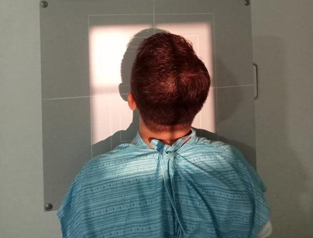
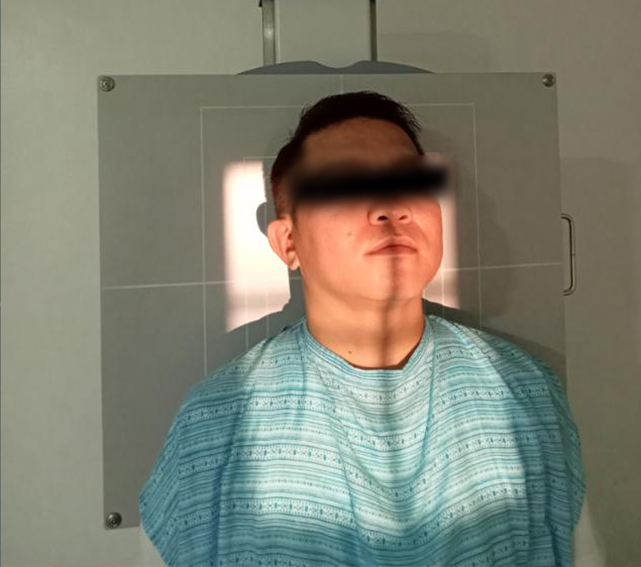
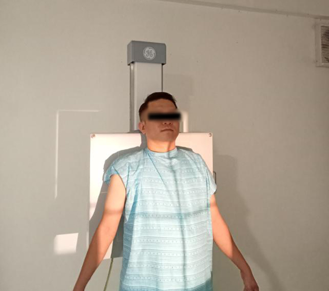
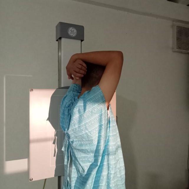
SKULL AP/L: Anteroposterior and lateral views of the skull to assess bone integrity and pathology.
NASAL BONE: Imaging of the nasal bones to evaluate for fractures or deformities.
MANDIBLE: Examination of the lower jaw to diagnose fractures or lesions.
MANDIBLE: Imaging of the eye socket to check for fractures or foreign bodies.
MASTOID: Evaluation of the mastoid process for infections or tumors.
NASAL AP: Anteroposterior view of the nasal area for detailed assessment.

Cervical and Thoracic Imaging
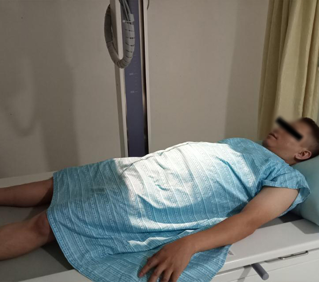
This imaging focuses on the cervical spine, thoracic spine, and chest structures. It is essential for diagnosing spinal conditions, assessing soft tissue, and evaluating lung health.
Services:
CERVICAL AP/LATERAL: Views of the cervical spine to assess alignment and fractures.
SOFT TISSUE AP/LATERAL: Imaging of neck and chest soft tissues to identify abnormalities.
LATERAL EXTENSION/FLEXION: X-rays of the cervical spine to evaluate motion and stability.
THORACIC VERTEBRA AP LAT: Views of the thoracic spine for pathology and alignment assessment.
CHEST PA/L (ADULT): Chest X-rays to evaluate lung fields and heart size.
CXR AP/L CHILD (0-15): Pediatric chest X-rays to assess respiratory and cardiovascular conditions.
Upper Extremity Imaging
This section covers imaging of the upper extremities, including the shoulders, arms, and hands. It aids in diagnosing fractures, joint issues, and other conditions affecting the upper limbs.
Services:
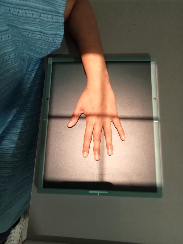
HAND PA/O/L: Posteroanterior, oblique, and lateral views of the hand to identify fractures and joint issues.
HUMERUS AP/L: Anteroposterior and lateral views of the humerus to evaluate fractures and tumors.
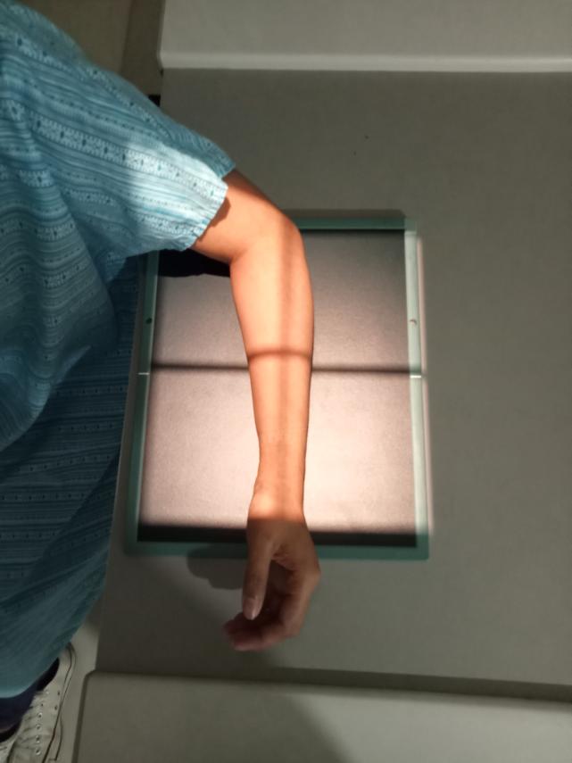
FOREARM AP/L: Anteroposterior and lateral views of the forearm to check for fractures or injuries.
ELBOW AP/O/L: Views of the elbow to assess joint integrity and fractures.
SHOULDER AP: Anteroposterior view of the shoulder to evaluate joint space and potential dislocations.
CLAVICLE: X-ray of the clavicle to detect fractures or misalignments.
Lower Extremity and Abdomen Imaging
This imaging area focuses on the lower extremities and abdominal region. It is essential for diagnosing fractures, joint conditions, and abdominal issues.
Services:FOOT AP/O/LAT: Posteroanterior, oblique, and lateral views of the foot to assess for fractures and structural issues.
ANKLE AP,MOT/L: Anteroposterior and lateral views of the ankle to evaluate injuries and joint alignment.
THIGH/FEMUR AP/LAT: Views of the femur to assess for fractures and other abnormalities.
LEG: X-ray of the leg to identify fractures or bone lesions.
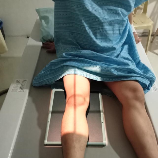
KNEE AP/LAT: Anteroposterior and lateral views of the knee to evaluate joint space and injuries.
TIBIA, FIBULA/AP/L: X-rays of the tibia and fibula to check for fractures and alignment issues.
PELVIS AP: Anteroposterior view of the pelvis to assess for fractures and joint integrity.
PELVIS FROGLEG: Special view to evaluate the hip joints and alignment with legs in a flexed position.
ABDOMEN UPRIGHT/SUPINE: X-rays of the abdomen to assess organ size and detect any fluid or gas accumulation.
APICO LORDOTIC: View targeting the apices of the lungs to identify lesions or infections.
STERNUM: X-ray of the sternum to assess for fractures or other abnormalities.